Menu
Menu


Despite a landscape clouded in complexity, investigational biomarkers are expanding our view of the pathophysiology of gastric cancer, and biomarker testing could provide a more comprehensive understanding of this disease.


CLDN18.2=claudin 18.2; FGFR2b=fibroblast growth factor receptor 2b; HER2=human epidermal growth factor receptor 2; MSI=microsatellite instability; PD-L1=programmed death-ligand 1.
INTRO
In Canada, estimates indicate that age-standardized 5-year net survival of patients diagnosed with stomach cancer is 25% (2008–2010).*,1
* Age-standardized 5-year net survival estimates for primary sites of cancer, both sexes, three years combined.

IN CANADA
Among patients who survive at least one year after diagnosis, approximately 55% survive for 5 years post-diagnosis (2015–2017).†,2
55% survive for 5 years post-diagnosis
In 2022, it was estimated that 4100 new cases of stomach cancers would be diagnosed in Canada (excluding Quebec).3
4100 NEW CASES
† Ages 15–99 years in Canada (excluding Quebec).
AROUND THE WORLD
An estimated 769,000 people died worldwide in 2020 due to G/GEJ cancers, making it the 4th most deadly cancer.4
Over 1 million new cases of G/GEJ cancers were diagnosed worldwide in 2020, making it the 5th most diagnosed cancer.4
In 2018, the global overall 5-year survival rate was approximately ≤10% in mGC.5
INVESTIGATIONAL BIOMARKERS
KNOWN BIOMARKERS
Investigational and known biomarkers can be detected by standard IHC staining methods.
INVESTIGATIONAL BIOMARKERS
CLDN18.2:
IHC8
FGFR2b:
IHC, ctDNA§,11,15
§ FGFR2b overexpression can be determined by IHC; FGFR2 gene amplification can be determined by ctDNA.11,15
KNOWN BIOMARKERS
PD-L1:
IHC||,6
HER2:
IHC, ISH, NGS¶,6,16
MSI/MMR:
PCR/NGS, IHC6
ctDNA=circulating tumour DNA; IHC=immunohistochemistry; ISH=in situ hybridization; MMR=mismatch repair; NGS=next generation sequencing; PCR=polymerase chain reaction.
|| Varying diagnostic assays.17
¶ Other ISH methods (CISH=chromogenic ISH; DDISH=dual-color dual-hapten ISH; FISH=fluorescent ISH; SISH=silver ISH).16
Investigational biomarkers are prevalent among mG/GEJ biomarkers.
Biomarker prevalence estimates from select international studies are reported below. Prevalence data can vary among studies due to tumour heterogeneity, differences in patient population, clinical trial methodology, and diagnostic assays used.8,12,18-21
INVESTIGATIONAL BIOMARKERS
CLDN18.28
(high expression)
29% primary cases
34% nodal metastases
FGFR2b18
(overexpressed)#
3–61%
# Overexpression determined by IHC.18
KNOWN BIOMARKERS
PD-L119,20
(Variable due to multiple factors)**
CPS ≥1: 67–73%
CPS ≥5: 29–31%
CPS ≥10: 16–18%
HER213
(Positive)
22%
MSI21
(MSI-high)
4%
CPS=combined positive score.
** Prevalence of PD-L1 at various CPS thresholds was explored.19

CLDN18.2
Claudins are a family of transmembrane proteins:9,22
Claudins are a major component of epithelial and endothelial tight junctions, which are involved in controlling the flow of molecules between cells.7-9
Claudins are present throughout the body, but two specific isoforms of CLDN18 are localized to certain tissue types:9,22
Preclinical studies have shown that CLDN18.2 may be exposed in gastric tumours.9
CONFINED IN
HEALTHY TISSUE


In gastric epithelial cells, CLDN18.2 is typically buried in the tight junction supramolecular complex.8,9
It functions to regulate selective barrier properties and contribute to cell-to-cell epithelial adhesion.8,9
RETAINED DURING TRANSFORMATION


The presence of CLDN18.2 is retained throughout malignant transformation, both in the primary tumour site and metastatic disease.23
CLDN18.2 may be expressed when tumours develop in pancreatic, lung, and ovarian tissues as well.9,23
An international study indicated that approximately 62% of primary gastric cancers and 55% of nodal metastases expressed CLDN18.2 (at any level), and approximately 29% and 34% of patients are CLDN18.2 positive (high expression), respectively.*,8
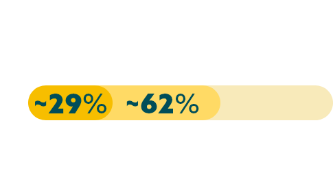
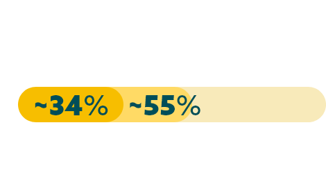
* Study population was limited to 523 patients with gastric (n=408) and gastro-esophageal (n=115) cancers, of which 195 patients had high expression of CLDN18.2.8
When evaluating the relationship between CLDN18.2 and other biomarkers, data from an international study suggests there is limited overlap.
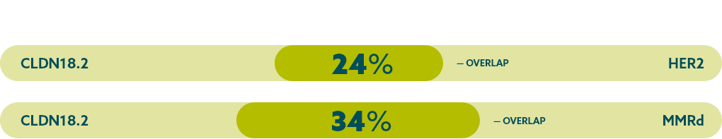
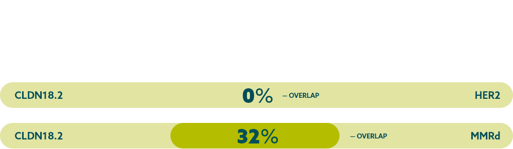
* Study population was limited to 523 patients with gastric (n=408) and gastro-esophageal (n=115) cancers, of which 195 patients had high expression of CLDN18.2. FGFR2b was not evaluated in this study.8
MMRd=mismatch repair deficient
CLDN18.2 is expressed in both diffuse-type tumours and intestinal-type tumours.8

FGFR2b
FGFR signaling mediates mitogenesis, differentiation, cell proliferation, angiogenesis and invasion.10
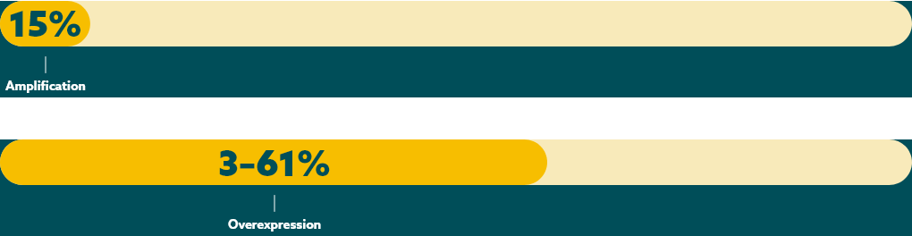
FGFR2b overexpression has been reported in up to 15%, and positivity has been observed in 3–61%, of mG/GEJ cancers.*,18 These data were reported in an international study.
* FGFR2b positivity: overexpression (IHC) and/or gene amplification.
Detecting FGFR2b overexpression can be done with the following tests.11,15

HER2
HER2 (human epidermal growth factor receptor 2) is a receptor-tyrosine kinase that is overexpressed and/or amplified in mG/GEJ cancer.6,10
HER2 positivity has been reported in an international study in
22% of advanced G/GEJ cancers.12


HER2 positivity: overexpression (IHC3+) and/or gene amplification (FISH positive)
Detection of HER2 may be done with IHC, ISH methods and NGS, and is generally more associated with the intestinal type.*,6,12,16
* IHC/ISH should be considered first, followed by additional NGS testing as appropriate.6

MSI
MSI is associated with genomic instability and increased susceptibility to tumour development.10
Microsatellites are repeated sequences of nucleotides in DNA.13
MSI-H has been reported in an international study in 4% of mG/GEJ cancers.21


Detection of MSI is typically assessed with various methods.6

PD-L1
PD-L1 (programmed death-ligand 1) is a transmembrane protein that may be expressed on various tumour cells and/or immune cells.28
Prevalence of PD-L1 has been reported for several positivity thresholds throughout various international studies: 67–73% CPS ≥1, 29–31% CPS ≥5, and 16–18% CPS ≥10.*,†,19,20


* Study population was limited to 592 patients with locally advanced or metastatic mG/GEJ cancer who experienced disease progression after first-line therapy with a platinum and fluoropyrimidine.20
† Study analyzed 56 specimens from therapy-naïve biopsies or post-neoadjuvant treatment from German patients with primarily non-metastatic gastric adenocarcinoma.19
PD-L1 expression is detected using IHC.6
SUMMARY
NCCN Clinical Practice Guidelines in Oncology for Gastric Cancer support biomarker testing.6
The NCCN Guidelines recommend:
Biomarker testing provides more insight into the pathophysiology of mG/GEJ cancer as more biomarkers are discovered.
* IHC/ISH should be considered first, followed by additional NGS testing as appropriate.6
As biomarker research continues, it expands our view of the patient population and reveals more information about the pathophysiology of mG/GEJ cancer.
REFERENCES
The information provided on gastriccancerbiomarkers.ca is intended for residents of Canada.
© 2023 Astellas Pharma Canada, Inc. All rights reserved.
WEBSITE TERMS OF USE (Effective date: 04/23)
ATTENTION: PLEASE READ THESE TERMS CAREFULLY BEFORE USING THIS WEBSITE GASTRICCANCERBIOMARKERS.CA. BY USING THIS SITE, INCLUDING ANY SERVICES PROVIDED ON OR THROUGH THIS SITE GASTRICCANCERBIOMARKERS.CA (“Services”), YOU INDICATE THAT YOU ACCEPT THESE TERMS. IF YOU DO NOT ACCEPT THESE TERMS, DO NOT USE THIS SITE.
Astellas Pharma Canada, Inc., (Astellas) may at any time revise these Terms of Use by updating this posting, so you should periodically examine the current Terms of Use to which you are bound. Certain provisions of these Terms of Use may be superseded by expressly designated legal notices or terms located on particular pages at this Site. To view the Terms of Use at any time, go to GastricCancerBiomarkers.ca.
GENERAL INFORMATION. This Site contains information that may be of interest to members of the healthcare community. Please feel free to browse this Site and Services. Your access and use of the Site are subject to the following terms and conditions, and all applicable laws. By accessing and browsing this Site and Services, you accept, without limitation or qualification, these terms and conditions and acknowledge that they supersede any other agreement between you and Astellas.
PRIVACY. Astellas policies concerning the use of your personal information are set forth in Astellas's Privacy Policy, which you can find by clicking here, and which are incorporated by reference herein. By using this Site or any services provided by or through this Site (Services), you agree to waive and release Astellas from any claim or liability in connection with the collection, use, or disclosure of information consistent with Astellas's Privacy Policy.
SITE CONTENT. All material on the Site including, but not limited to, text, images, graphics, documents, audio, or video (collectively, the Site Content) are protected under applicable copyright laws. You may copy, download, or print Site Content for your personal, noncommercial purposes only, but no modification or further reproduction of content is permitted. The Site Content may not otherwise be copied, downloaded, reproduced, deleted, modified, republished, retransmitted, displayed, or used to create derivative works without the express written permission of Astellas.
Trademarks, service marks, trade dress, logos, designs, and slogans appearing on this Site are owned by Astellas, its Affiliate companies, its licensors, or its partners, and may not be used for advertising or publicity purposes, or to indicate any affiliation with or endorsement by Astellas, except with the express written permission of Astellas.
Nothing contained in this Site should be construed as granting a licence to, or right in, any trademarks, patents, or copyrights of Astellas, or any other party.
You are solely responsible for any content, including but not limited to text, photographs, caricatures, illustrations, designs, icons, articles, audio clips, and video clips (collectively, User Content) that you post, email, or otherwise transmit via this Site. Astellas does not own or endorse User Content posted or otherwise transmitted via this Site, and Astellas does not guarantee the accuracy, integrity, or quality of such User Content. Any communications you send via this Site or otherwise to Astellas by email, with the exception of personally identifiable information as defined in Astellas' Privacy Policy, shall be deemed to be nonconfidential and Astellas shall have no obligation of any kind with respect to such information and shall be free to reproduce, use, disclose, and distribute the information to others without limitation. Astellas shall be free to use any ideas, concepts, know-how, or techniques contained in such information for any purpose whatsoever, including but not limited to developing, manufacturing, and marketing activities incorporating such information.
The content or material Astellas provides through the Site is for informational purposes only. Nothing on this Site should be construed as the giving of advice (medical, legal, financial, investment, or other professional advice) or the making of a recommendation, and information on the Site should not be relied on as the basis for any decision or action. This Site is not intended to promote any product or treatment, or to impact or influence any purchasing, prescribing or diagnosis decisions.
OBLIGATIONS OF SITE VISITORS OR USERS. You agree not to do any of the following while visiting or using the Site or any services provided by or through this Site:
Astellas may, in its sole discretion, take any action it deems necessary, including but not limited to termination of access to the Site or modification of User Content, if Astellas believes a user's conduct or content fails to conform to these Terms of Use or may create liability for Astellas, its partners, its suppliers, or any other party or person. Astellas will not be liable to you or any third party as a result of such termination or modification. The terms and conditions provided in these Terms of Use will survive any such termination and modification.
LINKS TO OTHER WEBSITES. Solely as a convenience to you, Astellas may provide links on this Site to other websites that are not maintained by or under the control of Astellas. If you use these links, you will leave this Site. Please remember that linked sites are not necessarily Astellas's sites, and the information could change without Astellas's knowledge. Third-party linked sites and their contents should not be attributed to Astellas (or any of its affiliated companies) in any manner. Astellas has not attempted to verify the truth or accuracy of any third-party sites or content contained on any third-party sites and does not make any representations or warranties about such sites or the content of such sites. If you decide to access any of the third-party sites linked to this Site, you do so entirely at your own risk. In the event Astellas endorses a particular organization, Astellas will clearly state its endorsement next to any link to that organization.
DISCLAIMER OF WARRANTIES. THIS SITE, INCLUDING ANY INFORMATION, SERVICES, OR MATERIAL MADE AVAILABLE ON OR THROUGH THIS SITE, IS PROVIDED ON AN "AS IS" AND "AS AVAILABLE" BASIS. TO THE MAXIMUM EXTENT PERMITTED BY LAW, ASTELLAS EXPRESSLY DISCLAIMS ALL WARRANTIES OF ANY KIND, EXPRESS OR IMPLIED, INCLUDING WITHOUT LIMITATION ANY WARRANTY OF MERCHANTABILITY, FITNESS FOR A PARTICULAR PURPOSE, OR NONINFRINGEMENT. FURTHERMORE, ASTELLAS DOES NOT WARRANT THE ACCURACY OR COMPLETENESS OF ANY INFORMATION ON THIS SITE AND MAY MAKE CHANGES TO THE INFORMATION, SERVICES, OR MATERIAL MADE AVAILABLE ON OR THROUGH THIS SITE AT ANY TIME WITHOUT PRIOR NOTICE TO USERS OF THIS SITE. INFORMATION AT THIS SITE THAT IS PERIODICALLY UPDATED MAY NOT BE CURRENT AT THE MOMENT YOU VISIT THIS SITE AND MAY CONTAIN ERRORS. ASTELLAS MAKES NO WARRANTY THAT THE SITE WILL MEET YOUR REQUIREMENTS; THAT THE SITE WILL BE UNINTERRUPTED, TIMELY, SECURE, OR ERROR-FREE; THAT MESSAGES OR REQUESTS WILL BE DELIVERED; THAT DEFECTS WILL BE CORRECTED; OR THAT THIS SITE IS FREE OF VIRUSES OR OTHER HARMFUL COMPONENTS. ASTELLAS MAKES NO WARRANTY REGARDING ANY GOODS OR SERVICES PURCHASED OR OBTAINED THROUGH THE SITE OR ANY TRANSACTIONS ENTERED INTO THROUGH THE SITE. YOU ASSUME THE ENTIRE RISK AS TO THE RESULTS AND PERFORMANCE OF THE SITE AND ANY GOODS OR SERVICES PURCHASED THEREFROM.
LIMITATION OF LIABILITY. IN NO EVENT WILL ASTELLAS BE LIABLE FOR ANY DAMAGES WHATSOEVER. THIS INCLUDES BUT IS NOT LIMITED TO DIRECT, INDIRECT, INCIDENTAL, PUNITIVE, AND CONSEQUENTIAL DAMAGES (INCLUDING, WITHOUT LIMITATION, THOSE RESULTING FROM LOST PROFITS, LOST DATA, OR BUSINESS INTERRUPTION) ARISING OUT OF THE USE, INABILITY TO USE, OR THE RESULT OF USE OF THIS SITE, ANY WEBSITES LINKED TO THIS SITE, THE MATERIALS OR INFORMATION CONTAINED ON ANY OR ALL SUCH SITES, OR ANY MATERIALS, PRODUCTS, OR SERVICES OFFERED ON THIS SITE OR SITES LINKED TO THIS SITE, WHETHER BASED ON WARRANTY, CONTRACT, TORT, OR ANY OTHER LEGAL THEORY AND WHETHER OR NOT ADVISED ON THE POSSIBILITY OF SUCH DAMAGES. IN NO EVENT WILL ASTELLAS, ITS SUPPLIERS, OR ANY OTHER PARTY INVOLVED IN CREATING, PRODUCING, OR DELIVERING THIS SITE BE LIABLE TO YOU IN ANY MANNER WHATSOEVER FOR ANY DECISION MADE OR ACTION OR NONACTION TAKEN BY YOU IN RELIANCE UPON INFORMATION PROVIDED THROUGH THIS SITE. APPLICABLE LAW MAY NOT ALLOW THE EXCLUSION OR LIMITATION OF INCIDENTAL OR CONSEQUENTIAL DAMAGES, SO THE ABOVE LIMITATION OR EXCLUSION MAY NOT APPLY TO YOU.
THIS SITE IS INTENDED FOR CANADIAN AUDIENCES ONLY. Access to and use of this Site is subject to the above terms and conditions. This Site was developed and is maintained by Astellas. Information, copy, and claims are intended only for the residents of Canada.